Once asked about medical history, family history and current symptoms, the healthcare provider may conduct a physical exam by palpating the upper right quadrant of the patient's abdomen and looking for a positive Murphy Sign. The provider may then order blood tests and imaging to be done. Some ways physicians can get images of potential gallstones are:
- Abdominal ultrasound
- Magnetic resonance cholangiopancreatography (MRCP)
- Hepatobiliary scintigraphy (HIDA) scan
- Abdominal CT scan
- Endoscopic retrograde cholangiopancreatography (ERCP)
- Cholecystogram
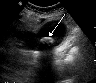
This image is an ultrasound depicting a large gallstone.
These tests are important in the diagnosis of cholelithiasis because they tell the provider if a gallstone is present, where it is, and if it is in a dangerous spot or not. The provider can then decide the best course of action.
"Bouveret Syndrome." Cinahl Information
Systems, Glendale, CA, 5 Dec. 2014. Web. 13 Oct. 2015.
Gallstones (Cholelithiasis). (2015, January 20).
Retrieved October 13, 2015.
Savitsky,
D. (14). Gallstones (M. Chwistek, Ed.). 20070420. Retrieved
September 9, 15, from Nursing Reference Center.

No comments:
Post a Comment